|
The 3 skin layers: epidermis, dermis, subcutaneous
fat
The skin is made up of three distinct
layers.
The top layer is called the
epidermis. (The word epidermis, and the name of the
other main skin layer, the dermis, both come from the name
used by the ancient Greeks for the skin, derma. From this we
also get the word dermatologist, meaning a doctor who
specialises in skin problems.)
The
epidermis is translucent. That is, it allows light to pass partially
through it, rather as frosted glass does. The epidermis does not
contain any blood vessels but gets its oxygen and nutrients from the
deeper layers of the skin.
At the bottom
of the epidermis is a very thin membrane, called the basement
membrane, which attaches the epidermis firmly, though not
rigidly, to the layer below.
The second
layer lies deeper and is called the dermis. It contains blood
vessels, nerves, hair roots and sweat
glands.
Below the dermis lies a layer of
fat, the subcutaneous fat. The depth of this layer differs
from one person to another. It contains larger blood vessels and
nerves, and is made up of clumps of fat-filled cells called
adipose cells.
The subcutaneous
fat lies on the muscles and bones, to which the whole skin structure
is attached by connective tissues. The attachment is quite loose, so
the skin can move fairly freely. If the subcutaneous tissues fill up
with too much fat the |
|
areas of attachment become more obvious
and the skin cannot move as easily -this is what gives rise to the
notorious cellulite (see pages 32 and
34).
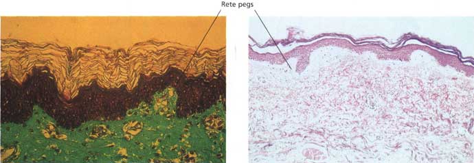
The junction between the epidermis and
the dermis is not straight but undulates like rolling hills - more
markedly so in some areas of the body than others. A series of
finger-like structures called rete pegs project up from the
dermis, and similar structures project down from the epidermis.
These projections increase the area of contact between the layers of
skin, and help to prevent the epidermis from being sheared off. They
are not present in the skins of unborn babies but rapidly develop
after birth, and are very noticeable in a young person's skin when
it is examined under the microscope. As skin ages they get smaller
and flatter.
Networks of tiny blood
vessels run through the rete pegs, bringing food, vitamins and
oxygen to the epidermis. In pale people these vessels can be seen
through the epidermis, particularly if the veins widen (so-called
'broken veins'). If the blood carries plenty of oxygen it will be
pink and the skin will tend to have a rosy color. If the blood is
running sluggishly and has lost most of its oxygen the skin will
look bluer. These blood vessels respond to temperature changes. They
open up in hot weather, bringing lots of red blood cells - and hence
a pink flush -to the skin, and close down in the cold; this is why
cold skin often looks blue.
(continued on next
page) |